- 541-884-3148
- info@klamatheyecenter.com
- 08:00AM to 5:00PM
Comprehensive Eye Exam
A complete eye examination does more than determine how clearly you see from a distance and which lens prescription, if necessary, will give you the best possible vision. Your eye doctor will also run a number of tests to check the health and function of your entire eye.
If you have never had an eye examination or are seeing a new eye doctor, your doctor or a technician will begin by asking you questions about your medical history, your family’s medical history, and any vision problems you may have. If you wear contact lenses, be sure to bring them with you to your appointment. Your eye doctor will check them to make sure that they are the correct prescription, fit, and kind of lens for your eyes.
A complete eye examination will include many or all of these painless tests:
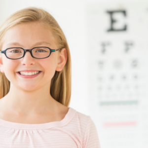
A visual acuity test measures how well you can see from a distance. Covering one eye at a time, you will look at an eye chart and be asked to identify letters that get smaller as you read farther down the chart.
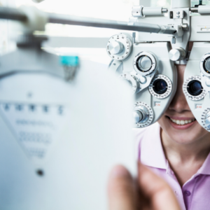
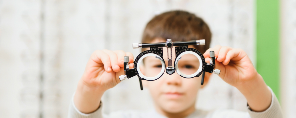
If your visual acuity test indicates that you need corrective lenses, you will be given a refraction test to determine the correct prescription. Your eye doctor may use retinoscopy to estimate your prescription by shining a light into your eyes to see the movement of the light reflected by your retina. The doctor will finalize your prescription by asking you to look through a device called a phoroptor that has many different lenses in it. You will be asked to compare a series of two lens choices and evaluate which lens combination provides you with your best possible vision correction.
To test the function of your eye muscles, your eye doctor will have you follow the movement of an object in many directions, looking for weak muscles or poor control of the muscles that move your eyes.
To test your peripheral vision, which is what you are able to see to the sides of your visual field when you look straight ahead, your doctor uses a visual field test. You may be asked to cover one eye at a time and, while looking straight ahead, tell your eye doctor when you can see his or her hand or other object as it moves inward from outside your visual field. Or a computer program may be used to test your visual field. If so, you will look straight ahead into a special device, often a lighted bowl-shaped instrument, and press a button each time you see a flash of light. Your eye doctor can use your responses to see if there are any blind spots in your visual field.
Your eye doctor will use a slit-lamp microscope to examine the front part of your eye, including the cornea, iris, and lens. You will sit at the slit lamp, which greatly magnifies your eye and shines a bright line of light into it, allowing your eye doctor to examine your eye closely. Before the test, you may be given eyedrops with fluorescein, an orange dye, to make your cornea easier to see. This dye will wash away naturally.
To test for glaucoma, a disease that can cause blindness when too much pressure in your eye damages the optic nerve, your eye doctor will use a tonometer to measure your intraocular pressure.
Using one method, noncontact tonometry, you will sit with your chin and forehead resting comfortably on the guides of a device that will blow a puff of air into your eye and thereby measure your eye pressure.
Applanation tonometry is another option. Your eye doctor will give you eyedrops containing an anesthetic and fluorescein dye to numb the front surface of your eye and will then use a manual tonometer to gently touch your cornea and measure the force required to flatten it. This procedure is quick and painless, and the anesthetic will wear off in 15 or 20 minutes.
Your eye doctor may also use pachymetry to measure the thickness of your cornea, which helps evaluate the accuracy of your intraocular pressure measurement. After applying numbing eyedrops, your ophthalmologist will use ultrasonic waves to measure your corneal thickness. This test is also a critical component of evaluating a patient’s candidacy for LASIK surgery.
A retinal examination explores the back of your eye including the retina and optic nerve. First, depending on the type of retinal examination your eye doctor chooses, your pupils will be dilated with eyedrops, which may sting briefly. If your eye doctor chooses to use direct examination, he or she will shine a light in your eye and use a device called an ophthalmoscope to look at the back of your eye. Alternatively, using a method called indirect examination, your eye doctor may use a much brighter light mounted on his or her forehead to examine your eye while holding it open. Finally, to get the best look at the back of the eye, your ophthalmologist may choose to perform a slit-lamp examination, which combines the use of the slit lamp and special lenses. Retinal examinations usually take about five minutes, but the eyedrops will continue to blur your vision for several hours. You may not be able to drive and will be sensitive to bright light, but this is temporary and should resolve in several hours.